Ewelina Tomecka
Warsaw University of Technology, Poland
Title: Nanofiber mats fabricated by solution blow spinning as potential substrates for cardiac cell culture
Biography
Biography: Ewelina Tomecka
Abstract
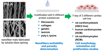
In recent years, there is a growing interest in the use of nanofibers for cell culture. Nano fibrous materials have many advantages such as: high porosity, high surface to volume ratio and they are structurally similar to extracellular matrix (ECM). Furthermore, nanofibers structure influence on cell orientation, which allow mimicking natural organization of cardiac cells. Most researches use electrospinning for nanofibers fabrication. However, low efficiency of this technique doesn’t allow to use it for large-scale production of nanofibers. We propose a rarely used method - solution blow spinning (SBS), which allows scale-up the nanofibers manufacturing process to the commercial level.This work presents the comparison and evaluation of cardiac cell proliferation on poly-(L-lactic acid) (PLLA) and polyurethane (PU) nano-fibrous mats fabricated by SBS. For the experiments, three different cardiac cell lines were used (figure 1).Cell cultures were performed for seven days on non-modified and protein-modified nanofibers surface. Obtained results of cardiac cell culture on investigated surfaces of nanofibers were compared to results of cardiac cell culture on polystyrene (PS) surfaces modified in the same way. The results showed the all types of investigated cells cultured on nanofibers (PLLA and PU) have more elongated shape than cells cultured on PS surface. Moreover, cells were arranged in parallel to each other, according to fibers orientation. In contrast, cells on PS surfaces were oriented randomly. Furthermore, in most cases, the cells proliferated better on nanofibers (PLLA and PU) than on PS surfaces modified in the same way. The results indicated that polymeric nanofibers (PLLA and PU) are better substrates for cardiac cell culture than PS surface and they enable cultivating these cells with conditions more similar to in vivo environment.

Figure. 1. Methodology of the experiments [5].
Recent Publications:
- Ricotti L, Polini A, Genchi G G, Ciofani G, Iandolo D, Vazão H, Mattoli V, Ferreira L, Menciassi A, Pisignano D (2012) Proliferation and skeletal myotube formation capability of C2C12 and H9c2 cells on isotropic and anisotropic electrospun nanofibrous PHB scaffolds. Biomedical Materials, 7: 035010.
- Orlova Y, Magome N, Liu L, Chen Y, Agladze K, (2011) Electrospun nanofibers as a tool for architecture control in engineered cardiac tissue. Biomaterials, 32: 5615–5624.
- Ashammakhi N, Ndreu A, Yang Y, Ylikuppila H, Nikkola L (2012) Nanofiber-based scaffoldsnfor tissue engineering. European Journal of Plastic Surgery, 35: 135 – 149.
- WojasiÅ„ski M, Pilarek M, Ciach T (2014) Comparative studies of electrospinning and solution blow spinning processes for the production of nanofibrous poly(L-lactic acid) materials for biomedical engineering. Pol. J. Chem. Technol. 16: 43–50.
- Tomecka E, Wojasinski M, Jastrzebska E, Chudy M, Ciach T, Brzozka Z (2017) Poly(L-lactic acid) and polyurethane nanofibers fabricated by solution blow spinning as potential substrates for cardiac cell culture. Materials Science and Engineering C 75: 305–316.

